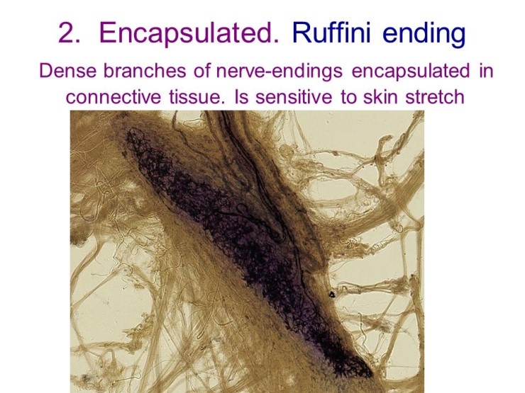

These are the structures at the terminals of Slowly Adapting Type I receptors. Branches of one axon makes contact with many Merkel cells which sense indentation of the skin. Merkel Cells exist in the basal layers of the epidermis and enclose the terminals of some large myelinated axons. Large axons also innervate the skin, and mediate the senses of Touch and Vibration. These four receptors allow for the sensation of joint movement, joint position and the extremes of movement associated with pain.
#Ruffini endings free
Golgi receptors and free nerve endings are also present around joints. Paciniform corpuscles are akin to the Pacinian Corpuscles found in other tissues, but have less lamellae in the onion-like encapsulation of the ending. Ruffini endings akin to the Type II slowly adapting receptors found in the dermis are present in the connective tissues around joints. Joint receptors are found in the synovial membranes, joint capsules and ligaments, and respond to mechanical changes in joint position, and play an important role in kinaesthesia and proprioception.įour types of receptor endings are recognised in the synovial membranes and connective tissue/ligaments around joints. These afferents are sometimes called Ib afferents. Golgi Tendon Organs arise from tendons and have large diameter axons that are slightly smaller than those of the muscle spindle. The latter are called gamma-efferent fibres and are finely myelinated. Other nerve fibres innervating the spindle include (a) a smaller diameter (group II) afferent and (b) some fibres that cause the intrafusal muscle fibres to contract. The largest afferent fibres are the Ia afferents arise from the a sensory ending that spirals around the central region of the intrafusal fibres- sometimes called the annulo-spiral ending. The muscles spindle has both afferent and efferent innervation. The structure is orientated in a parallel direction with the much larger extrafusal fibres, and is attached to the endomysium that surrounds the extrafusal fibres. has a bulge in its central region and is surrounded by a capsule throughout its length it contains modified muscle fibres called intrafusal (meaning inside the spindle) fibres. The Muscle Spindle is spindle shaped- i.e. Joint receptors are also involved in kinaesthesia. These provide the nervous system with information about muscle length and tension, and contibute to kinaesthesia - the sense of position, particularly in a limb. The largest afferent axons in peripheral nerves innervate muscle spindles and tendons.

Muscle Spindles and Golgi Tendon Organs Top In myelinated axons, the Schwann cell wraps itself around the axon many times, and the multiple layes of cell membrane act as the fatty insulator called myelin. multiple layers of cell membrane) is not formed around these axons. In these diagrams of peripheral nerves, several unmyelinated axons are invaginated into a Schwann Cell (forming a Remak Bundle) but myelin (i.e. The axons with the largest diameters have the highest degree of myelination These axons are often divided into groups depending on the degree of myelination: unmyelinated, finely myelinated and large myelinated. Neurones with cell bodies in the dorsal root ganglia have perikarya and axons of different diameters. The terminal branches of these neurones are unmyelinated and spread throught the dermis and into the epidermis of the skin, or around muscle fibres of skeletal muscle, or around blood vessels in the viscera.
#Ruffini endings skin
Encapsulated Nerve Endings : Muscle Spindles and Golgi Tendon Organs Sensory Receptors in Joints Encapsulated Endings in Skin : Merkel Cells, Meissner's Corpuscles, Ruffini end organs, Hair Receptors The Pacinian Corpuscle Free Nerve Endings and TRPV ion channels


 0 kommentar(er)
